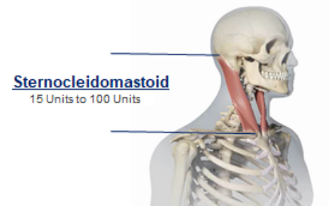
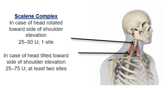
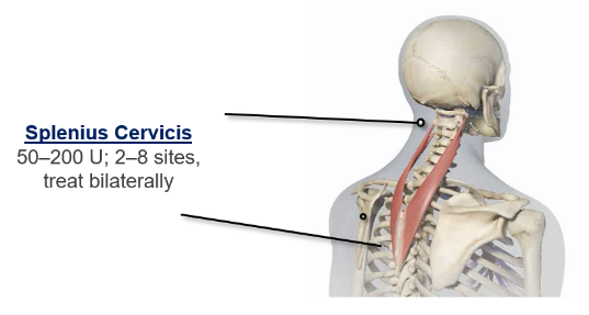
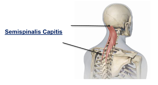
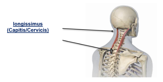
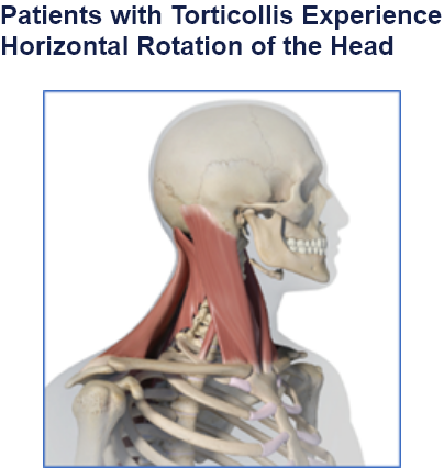
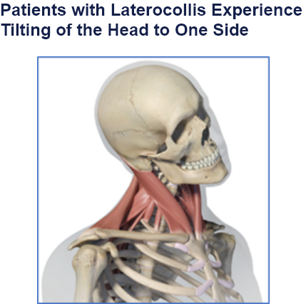
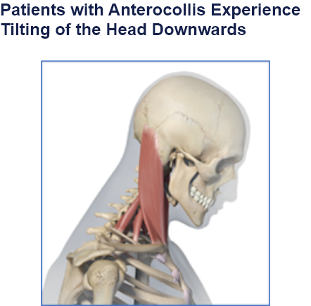
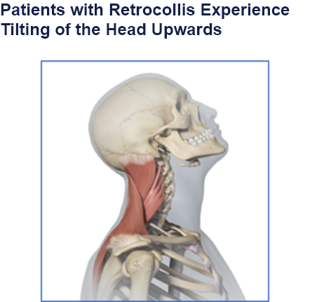
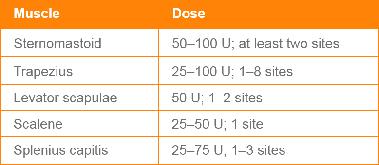
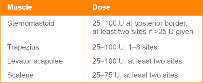
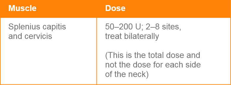
Clinical Presentation Pattern and Associated Muscles
Injection Sites
Patterns and Clinical Presentations
Botulinum Toxin Type A Dosing in Cervical Dystonia
Maximum total dose:
A total dose of 300 Units at any one sitting should not be exceeded. The optimal number of injection sites is dependent upon the size of the muscle. Treatment intervals of less than 10 weeks are not recommended.
Type I: Head Rotated Toward Side of Shoulder Elevation
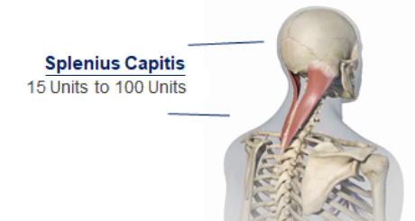
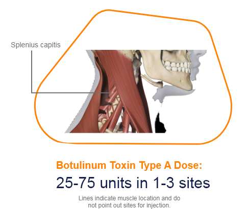
The splenius capitis is a strap-like muscle that, along with the splenius cervicis, comprises the superficial layer of intrinsic back muscles.1
• Function: The muscle flexes and rotates the head and neck to the same side; particularly in the superior and inferior lateral oblique movements.2
• Origin: Begin on the spinous processes of vertebrae from C-7 to T-3 and the ligamentum nuchae.2
• Insertion: The insertion extends from the medial edge of the mastoid process and the lateral part of the superior nuchal line.2
• Innervation: The nerve supply to the splenius capitis is provided by lateral branches of the posterior rami of the middle and lower cervical spinal nerves.2
.
Muscle Function Animation
.
.
Cadaver Demonstration
.
References
1. Gaillard, Frank. “Splenius Capitis Muscle: Radiology Reference Article.” Radiopaedia Blog RSS, 2 Aug. 2021. Available at: radiopaedia.org/articles/splenius-capitis-muscle. [Accessed 1 August 2023].
2. Ernest E, Ernest M. (2006) Splenius Capitis Muscle Syndrome. Pract Pain Manag.;6(5).
3. Bordoni, B. and Varacallo, M. (2018). Anatomy, head and neck, sternocleidomastoid muscle.
4. V, Deng F, A A, et al. Sternocleidomastoid muscle. Reference article, Radiopaedia.org (Accessed on 02 Aug 2023) Available from: https://radiopaedia.org/articles/13075
5. Ourieff, J., Scheckel, B. and Agarwal, A. (2018). Anatomy, Back, Trapezius.
6. Javed, O., Maldonado, K.A. and Ashmyan, R. (2022). Anatomy, Shoulder and Upper Limb, Muscles. In StatPearls. StatPearls Publishing.
7. Henry, J.P. and Munakomi, S. (2020). Anatomy, head and neck, levator scapulae muscles.


